
Exploring New Depths of Aquatic Health at the Mississippi Aquarium
By Kathleen Wendover, DVM ‘26
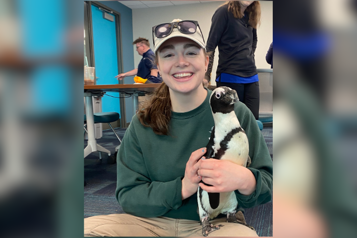
Earlier this year, I gained invaluable, hands-on experience at the Mississippi Aquarium in Gulfport, Mississippi. As my first externship in an aquarium setting, this experience opened my eyes not only to the diversity of species treated, but also to the breadth of diagnostic and treatment approaches available in an aquarium setting. My time at the Mississippi Aquarium allowed me to develop important connections as well as better appreciate the wide range of exciting paths my veterinary career may be able to take.
Adapting techniques to aquatic populations
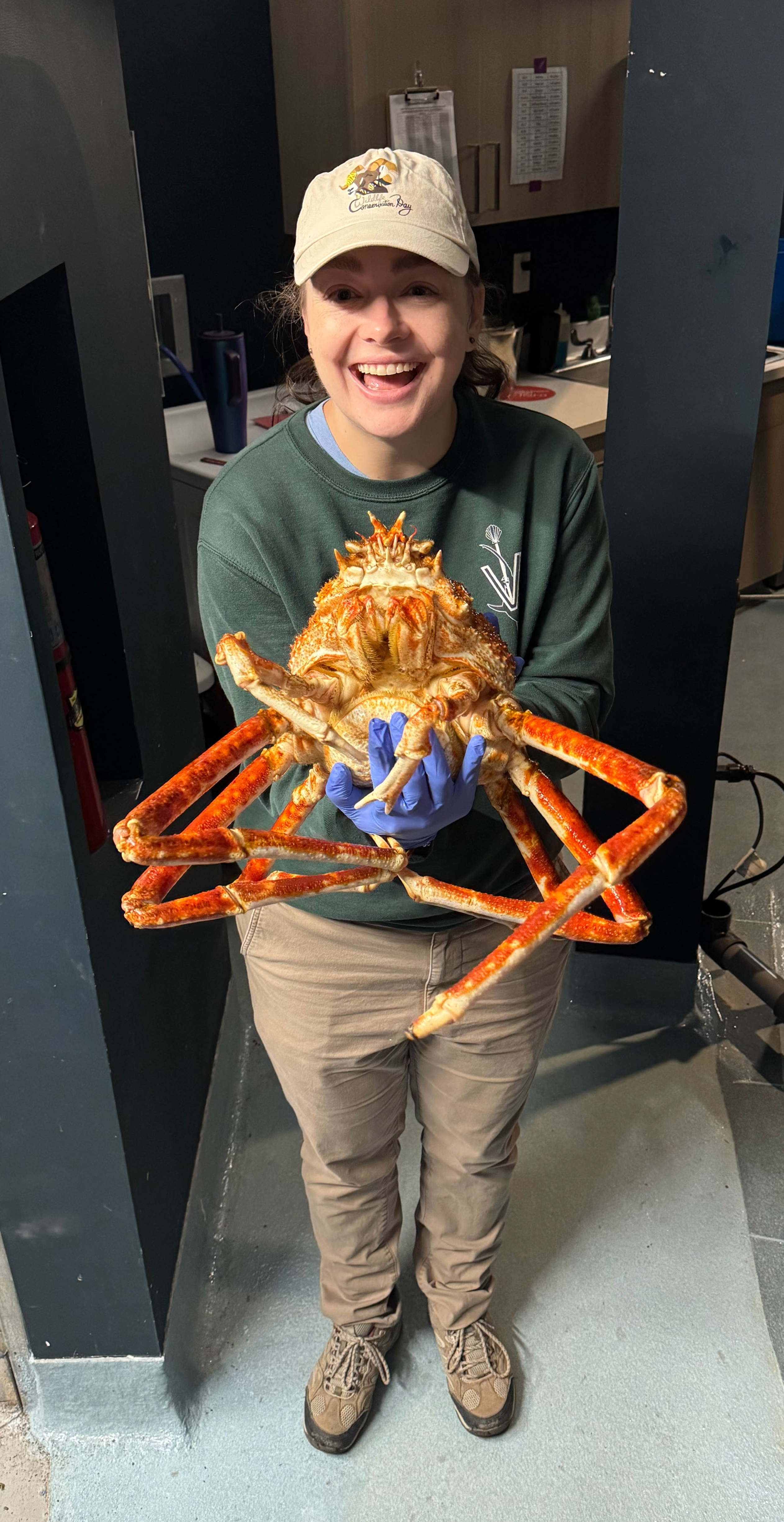
My externship at the Mississippi Aquarium allowed me to work with a variety of unique species, often for the first time. From a Japanese spider crab with a lesion on its limb to assessments of porcupine fish and river otters, my work with the veterinary team spanned multiple taxa. I assisted in the treatment of various invertebrate, amphibian, elasmobranch (cartilaginous fish), teleost (bony fish), reptilian, avian, and mammalian patients.
While a large portion of our cases were onsite at the aquarium, the veterinary team made frequent trips to the Aquatic Research Center (ARC), a separate non-exhibit facility. Animals at ARC may be under quarantine as new arrivals, off exhibit (e.g. African penguins), or undergoing rehabilitation (e.g. Kemp’s Ridley sea turtles). The scope of the clinical care performed by the veterinary team while I was there ranged from daily checks on injured or ill animals to procedures requiring anesthesia.
Participating in these cases allowed me to adapt clinical techniques I had learned previously, such as general physical examination, wound assessment, medication administration, and ophthalmologic evaluation, to aquatic patients. I was able to see diagnostics adapted to various species including voluntary gastroscopy in dolphins, ultrasounds on rays, and cloacal washes on fish. For cases that required it, I was able to hone my venipuncture skills collecting blood samples from sea turtles and different elasmobranchs.
Population dynamics at the aquarium
Consideration for population dynamics served as another factor during weekly routine checks and follow-ups on sick or injured animals. Because aquarium populations incorporate a variety of taxa into one tank, often these animals can experience predation or competition resulting in injury. As mixed populations are unavoidable in an aquarium setting, I saw a great deal of consideration go into relocating or adjusting populations in certain tanks to minimize problems.
Throughout my time at the aquarium, the veterinary team was monitoring the health and recovery of a porcupine fish named Benjamin who had a large wound on his tail. The wound was suspected to be from predation, and healed well with pain medication, antibiotics, and topical treatment. However, Benjamin’s reintroduction proved difficult with the looming threat of predation and competition for resources. After careful consideration and separation when necessary to heal, Benjamin was ultimately able to be integrated back freely into the tank. The population dynamics faced by multiple tanks gave me a newfound appreciation for the unique challenges that aquarium vets face while incorporating social structure into the overall health assessments of their patients.
Octocorals at the Aquatic Research Center
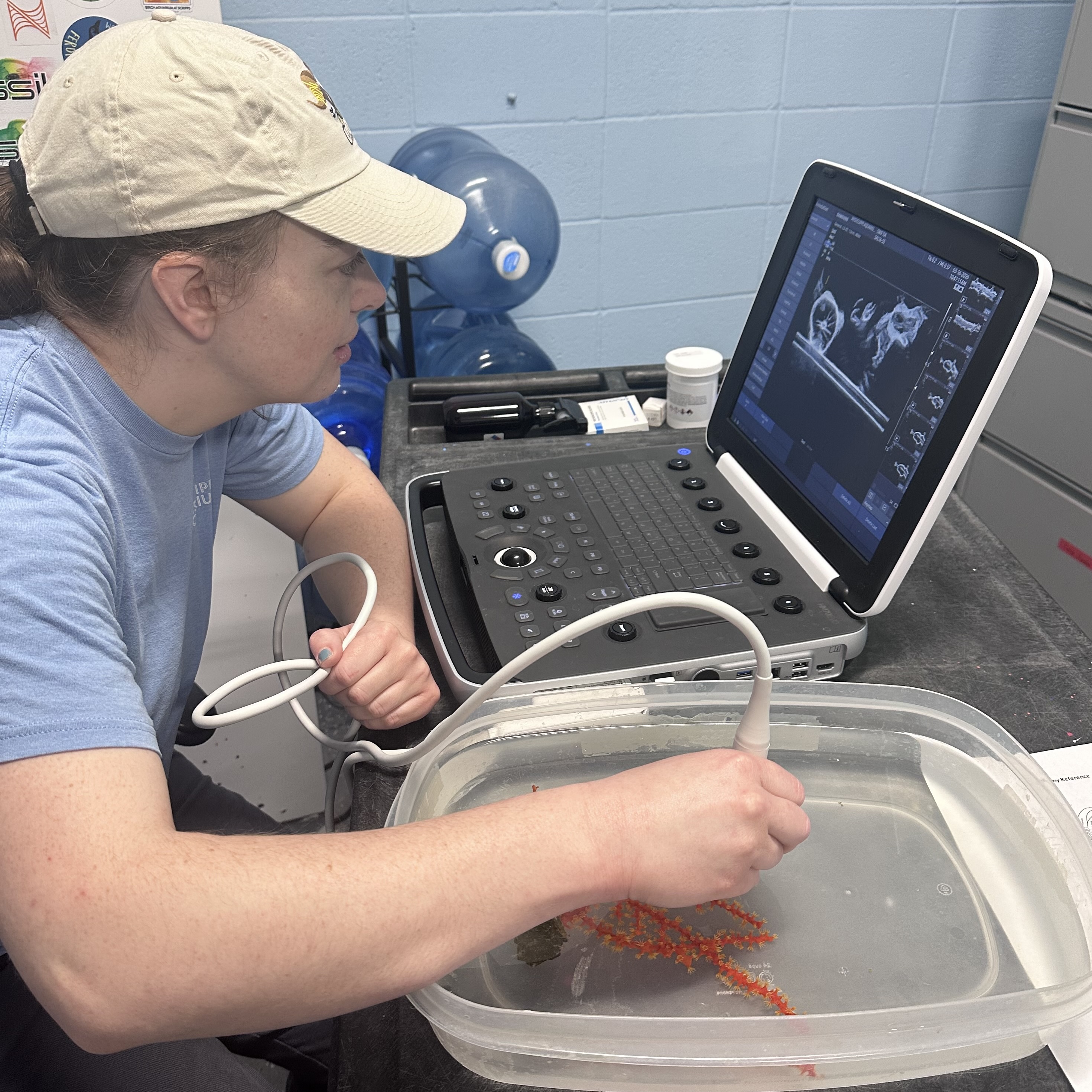
During my externship, I was able to spend time working on corals. The aquarium has a dedicated team based out of the ARC that does a lot of work with octocorals specifically. Although many of their octocorals were thriving, there was one population that continued to struggle due to acute changes in their tank’s water quality. We conducted scrape and impression cytology to screen for diseases on the surface of the corals. Additionally, I helped perform ultrasounds on corals with swollen branches, which were then aspirated to help investigate the fluid contained within the swellings.
While these diagnostics helped to clarify the extent of the disease in these corals, the exact cause for the pathology could not be determined. This prompted me to complete a literature review and presentation on the physiology and diseases of octocorals. This project gave me a greater appreciation for the complexity of coral physiology, the need for further coral research, and the challenges of maintaining coral health in aquaria.
Rehabilitation of Kemp’s Ridley sea turtle
An unforgettable highlight of this externship was aiding in the rehabilitation and release of Kemp’s Ridley sea turtles.
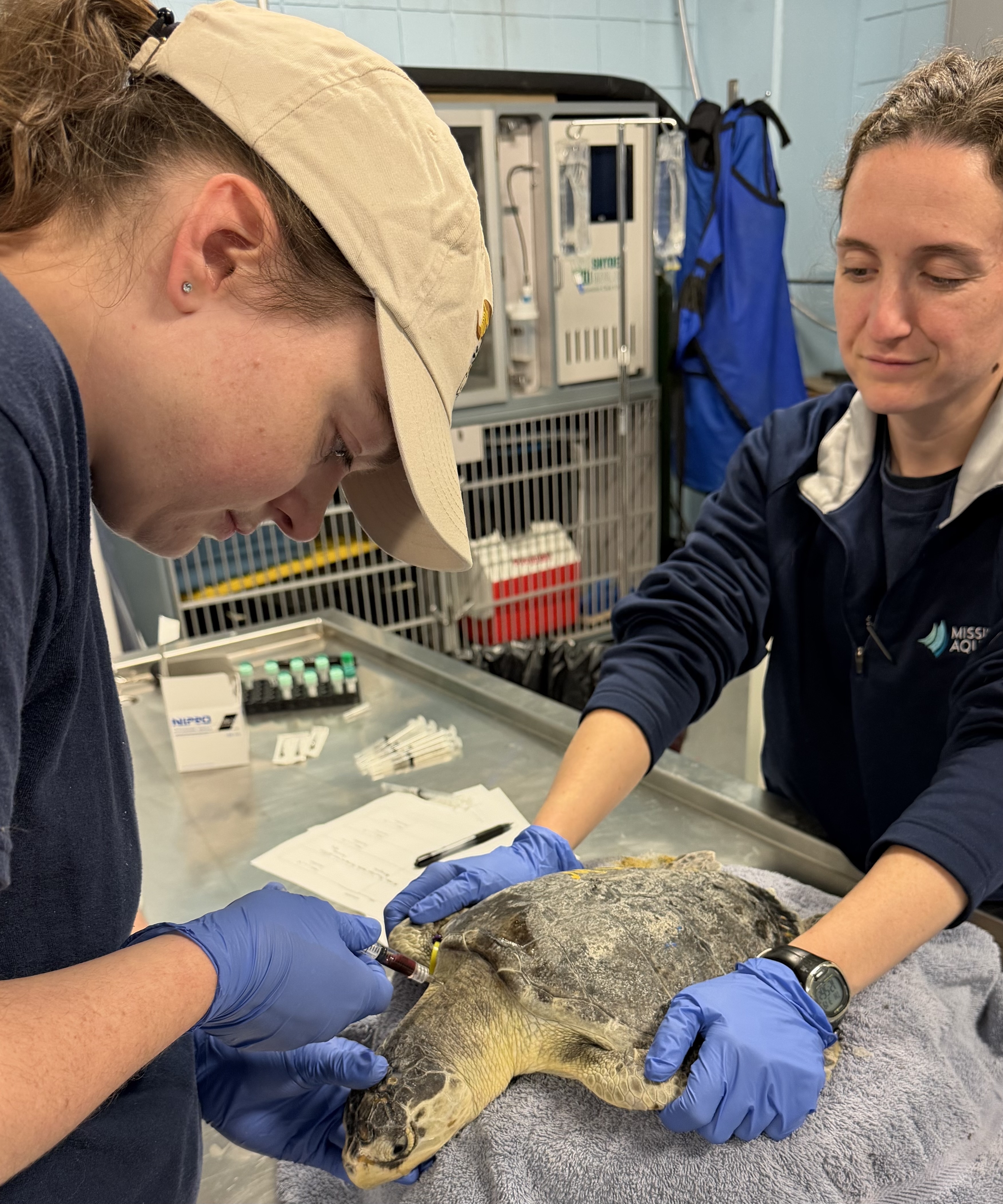
Most of these turtles were originally rescued in the fall of 2025 after being cold-stunned off the coast of Massachusetts. This unfortunately annual occurrence leaves many sea turtles in need of care over the winter as they recover from injury or illness. Mississippi Aquarium serves as one facility in a network of locations where these turtles can receive excellent care and medical attention to give them the best chance of survival.
With the timing of my externship in the spring, I participated in the release preparations for the recovered sea turtles, as well as the targeted care of those with more severe injury or illness. The turtles that recovered well received pre-release exams, bloodwork, and radiographs. During this time, I was able to gain practice examining the turtles for signs of illness or injury, collecting blood samples, and assisting with diagnostic imaging.
Diagnostic imaging served as a valuable tool for assessing illness among the Kemp’s Ridley sea turtles at ARC. Radiographs were largely used to assess lung health, as there was a general concern regarding pneumonia among turtles that were cold-stunned and had stranded. When turtles were not thriving or had significant lung lesions, they were taken to a local human hospital for CT scans. During my first week at the Mississippi Aquarium, I accompanied the veterinary team as several sea turtles underwent scanning. The CT scans informed next steps for the veterinary team: identifying turtles ready for release or individuals with lung pathology that required continued treatment.
As a rewarding addition to my time with the sea turtles, I participated in the release of 28 turtles. Taking part in the return of this critically endangered species to the wild served as an inspiring reminder of the goals I aim to accomplish in my career as a veterinarian. It also gave me a deeper appreciation for the impact that can be made when zoos and aquaria participate in wildlife conservation efforts. Learning about the network of institutions participating in the rehabilitation of these sea turtles increased my awareness of the threats faced by wild populations.
Conclusion
I am so grateful to Drs. Laken Russell and Alexa Delaune, as well as Kristen Hales, for teaching me so much during this experience. At the Mississippi Aquarium, I gained a wealth of information not only from the veterinary team, but also from the Ambassador, Aquatics, Dolphin, and Life Support teams. Throughout this externship, I developed a greater understanding of the wildlife health field and formed strong connections with dedicated individuals working to ensure the welfare and health of aquatic wildlife. Spending time with staff members from multiple teams allowed me to enhance my clinical reasoning, while broadening my range of clinical skills.
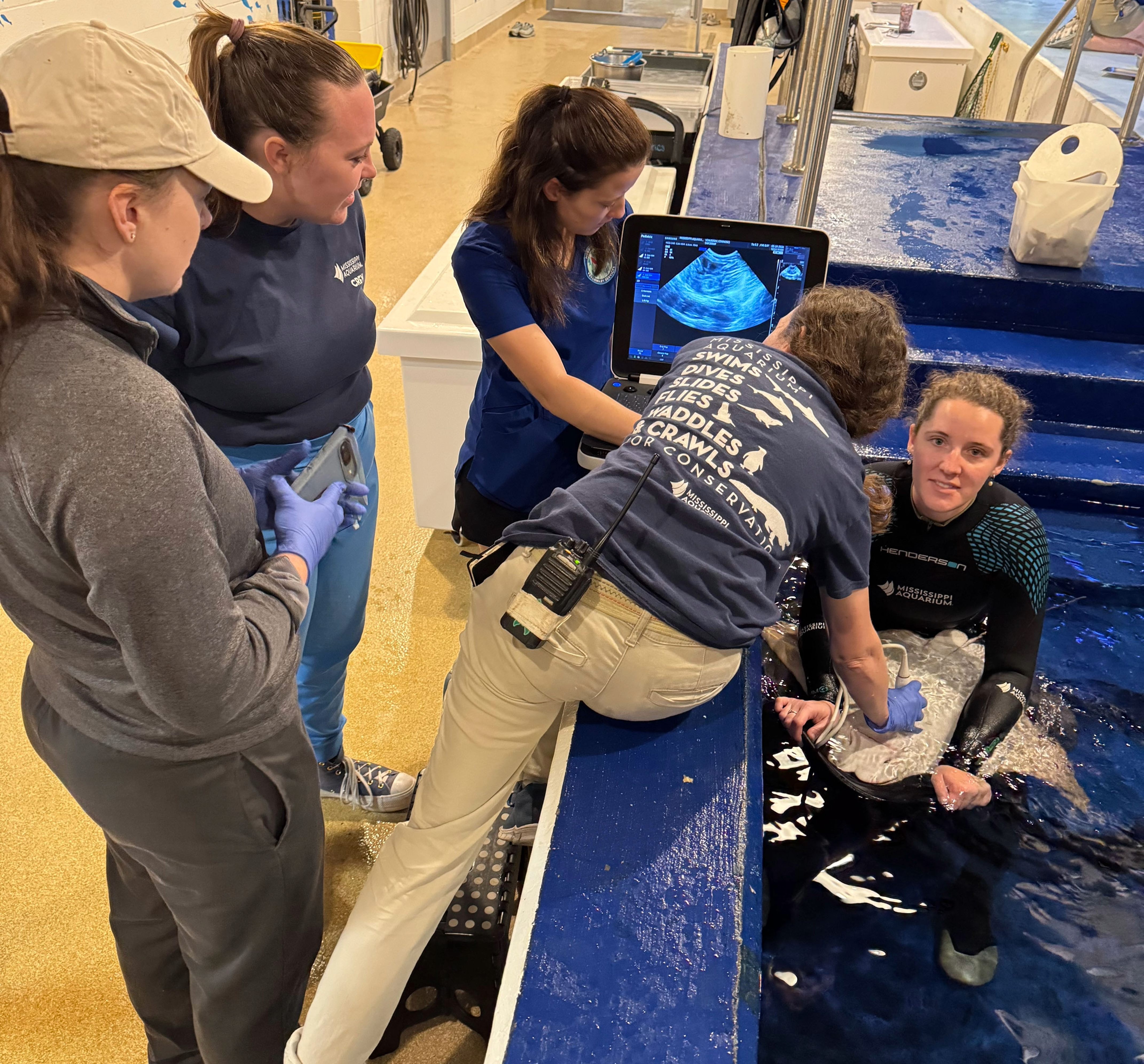
The opportunity to treat wildlife in a rehabilitation setting was extremely fulfilling and will continue to inspire me. I want to give special thanks to the Cornell K. Lisa Yang Center for Wildlife Health Student Support Fund for helping to make my clinical year extraordinary with such a significant experience.
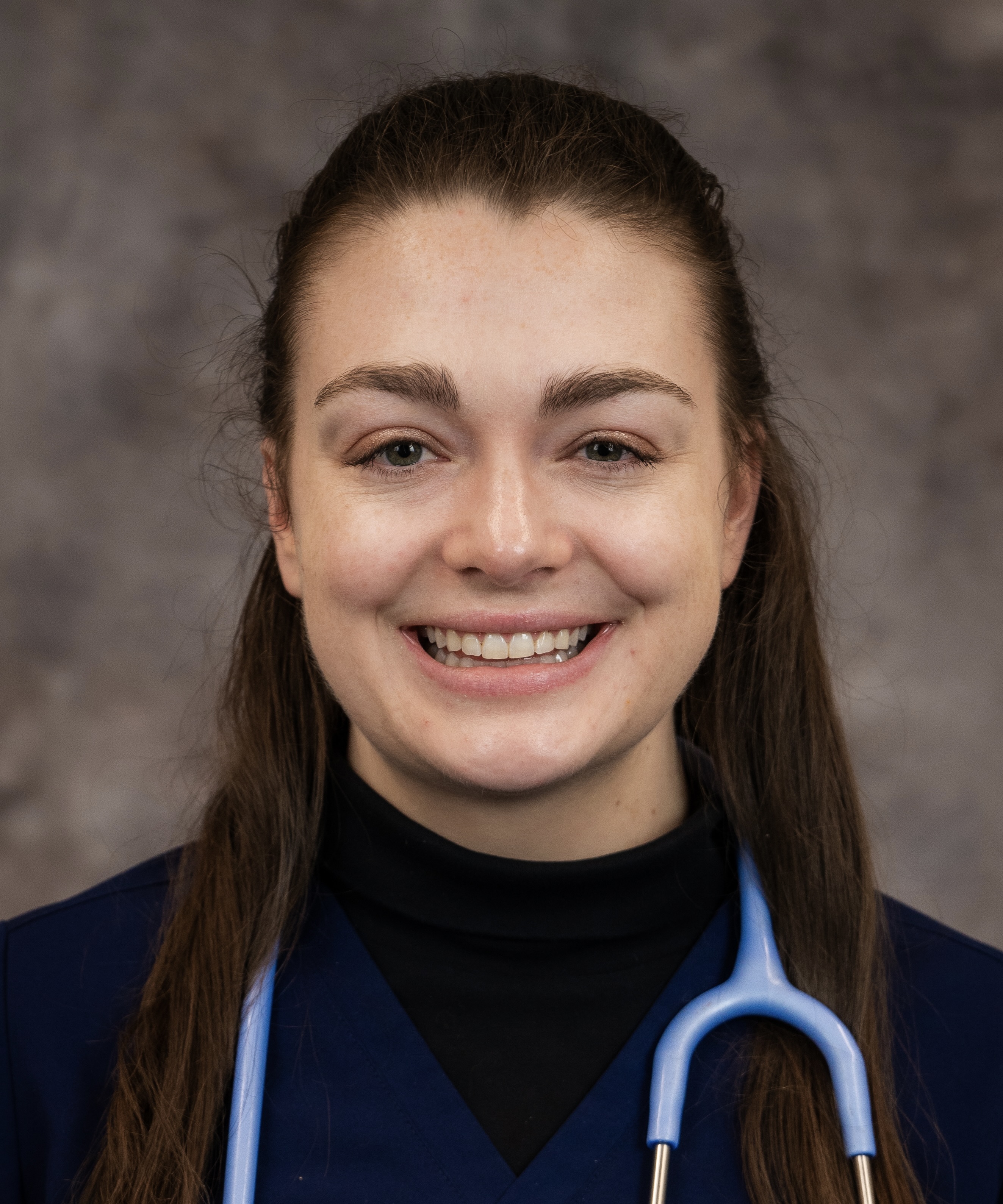
Kathleen Wendover, Class of 2026, is a recent graduate of the Cornell University College of Veterinary Medicine. She obtained a BA in biochemistry and molecular biology from Hendrix College in Conway, Arkansas. She is passionate about wildlife medicine, with a special interest in aquatics, and aims to pursue a career in wildlife conservation focused on population health.
All photos and videos provided by Kathleen Wendover unless otherwise noted.
Please consider giving to the Cornell Yang Center for Wildlife Health Student Support Fund to help provide more hands-on experiential learning opportunities for students passionate about wildlife health and conservation.