Using Veterinary Pathology to Investigate Marine Mammal Strandings in the North Sea
Every seal or porpoise that strands tells a story of the life it lived and how it died, though some findings from necropsies make more complete stories than others. When marine mammals, including seals and cetaceans, are found dead on the beach in the Netherlands or die at a rehabilitation center, members of the public and marine health experts contact the Strandings Investigation Program at Utrecht University’s Faculty of Veterinary Medicine. I spent four weeks as an extern with the program, gaining hands-on experience performing necropsies and learning more about wildlife health through the lens of pathology.
Free-ranging, healthy marine mammals are rarely captured for full veterinary examinations. When they are caught, the exams are brief due to the complex nature of providing anesthesia for these animals; limiting handling time reduces stress and allows them to return to the water as soon as possible. In Europe, a stranding is defined as when a dead or living marine mammal lies entirely on land in a helpless state, which may be due to illness, injury, or simply being lost. Live animals showing clear signs of physiological dysfunction in shallow waters would also be considered “stranded.”
While we don’t hope for an animal to become stranded, marine mammals that strand and succumb to their injuries provide conservationists with the unique opportunity to conduct a full postmortem examination. These investigations, or necropsies, include a thorough examination of all organs, microscopic analysis of tissues, blood chemistry tests, urinalysis, and infectious disease testing. While the findings cannot help that particular animal, they can be used to protect and improve the health of other animals in the future.
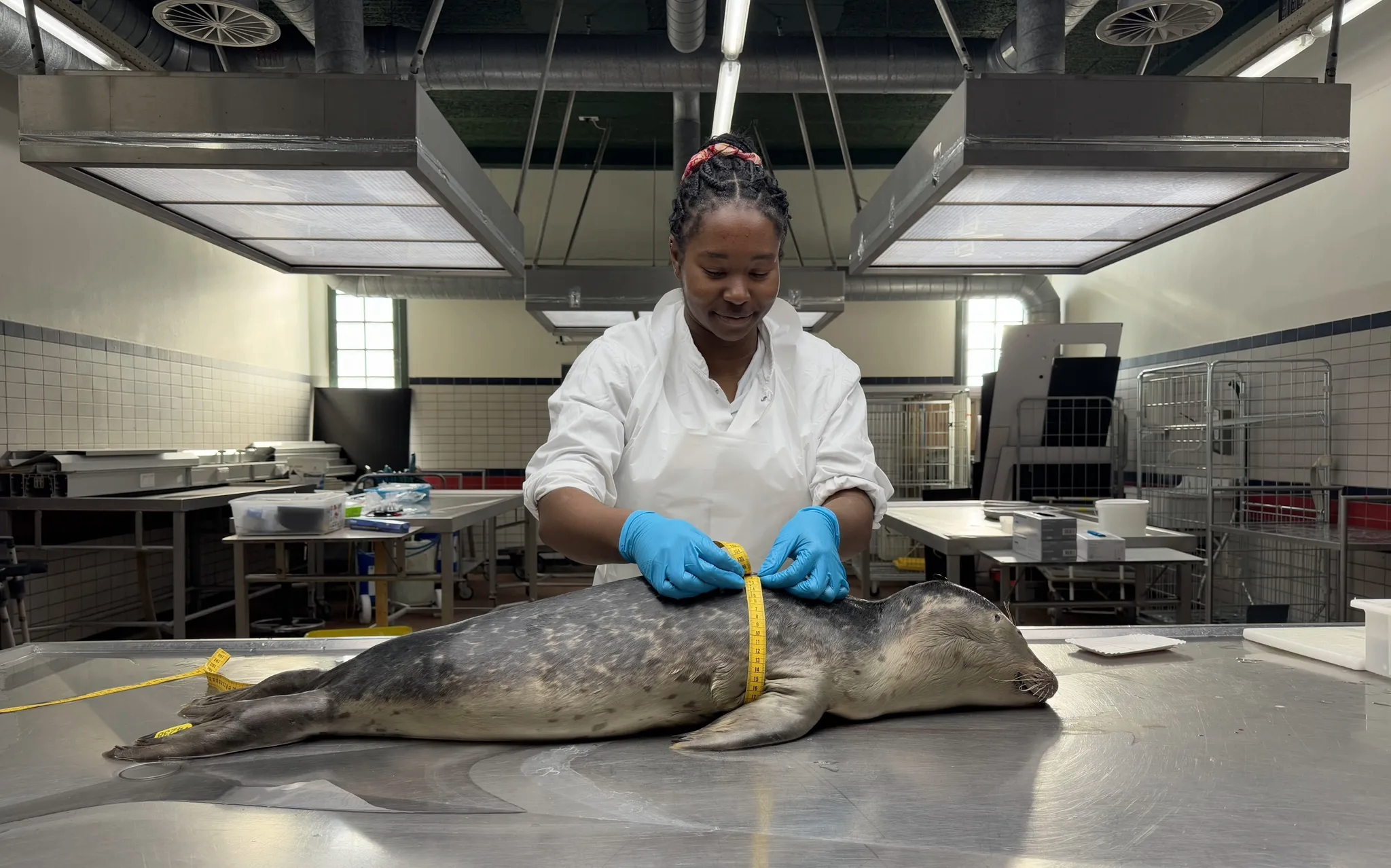
Each week in Utrecht, I participated in postmortem investigations or reviewed histopathologic cases from harbor porpoises (Phocoena phocoena), grey seals (Halichoerus grypus), and harbor seals (Phoca vitulina). Most animals that we examined were from the North Sea, a part of the Atlantic Ocean bordered by several European countries. This means that the Strandings Investigation Program may evaluate animals that have previously lived in waters near other parts of Europe. While I was there, we performed a necropsy on a seal that had tags from a rehabilitation center in Germany! I also joined the Dutch Wildlife Health Center for a postmortem examination of a Eurasian river otter (Lutra lutra).
The necropsies were great hands-on experiences. I learned how to assess individual organs and was able to discuss findings with board-certified veterinary pathologists and residents. I then further refined the morphologic descriptions and diagnoses while writing necropsy reports, which we completed for every animal. This training strengthened my ability to systematically evaluate each organ and clearly describe findings so that someone who was not present at the necropsy would be able to envision what was found.
If people contribute to threats facing wildlife, we should be equally—if not more—invested in finding solutions that can improve wildlife health and animals’ quality of life in the ocean.
One of the most memorable cases we investigated was a grey seal that had likely been entangled in a fishing net or other marine debris shortly before its death. It is awful to see evidence of human involvement in the deaths of these animals, but such cases also fuel my desire to pursue a career in wildlife health. If people contribute to threats facing wildlife, we should be equally—if not more—invested in finding solutions that can improve wildlife health and animals’ quality of life in the ocean.
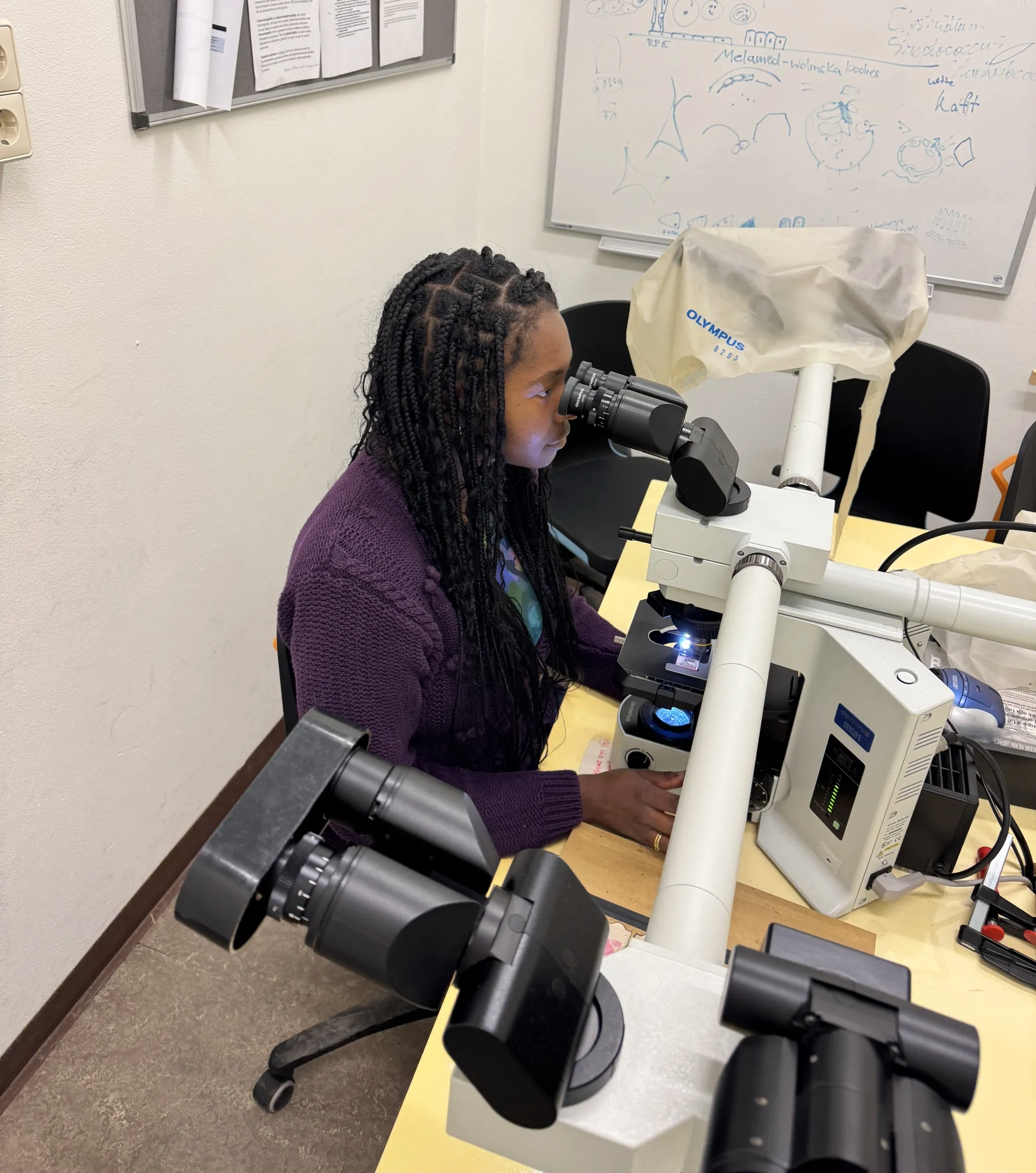
When not performing necropsies on gross specimens, I learned to recognize histopathologic features—abnormalities that can only be found by looking at tissues under a microscope—of diseases from past cases. Given a set of slides prepared from organs, I examined each organ’s slide and wrote down questions and observations. Then, I discussed my findings with the pathology resident and compared them to the official report written by a pathologist. This was invaluable for me, because I was able to show the resident exactly what I was looking at under the microscope and receive immediate, one-on-one feedback.
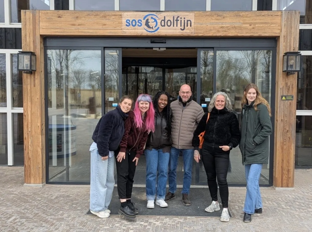
I also joined the Strandings Investigation Program on a visit to SOS Dolfijn, a marine mammal hospital and rehabilitation center about two hours north of Utrecht. Their facility includes indoor and outdoor pools for hospital patients, observation decks for staff, and a hospital suite. We were able to see staff members train two recently admitted seals to touch their noses to a target in order to receive a fish at mealtime. This training allows veterinary staff to visually assess patients multiple times per day.
I also joined a research project related to infectious disease in harbor porpoises. While helping with this project, I reviewed years of postmortem investigation reports, photos, and data. A single necropsy may not always yield clear results for a stranded animal, but this research project demonstrated to me that recognizing patterns across multiple cases can lead to significant discoveries regarding the health of wildlife populations. Disease research anchored in the analysis of pathologic lesions and patterns is important because, in the future, findings from historical data can help aquatic veterinarians recognize and treat diseases in live animals.
This externship deepened my passion for pathology as a means of understanding the health of marine mammals. Pathology is crucial for marine mammal health and conservation because it is one of the most effective ways to identify infectious disease outbreaks and monitor disease prevalence in populations that spend their entire lives in the ocean. I am extremely grateful to the pathologists who spent so much time teaching me one-on-one, especially Drs. Linde van Schalkwijk, Lonneke Ijsseldijk, and Nadiah van Eijk. I am also thankful to Dr. Jennifer Bloodgood for helping me organize this externship, and to the Cornell K. Lisa Yang Center for Wildlife Health Student Support Fund for funding this incredible learning experience.

Victoria Priester, Class of 2026, is a fourth-year veterinary student at Cornell University’s College of Veterinary Medicine. She is interested in free-ranging wildlife medicine and conservation, and enjoys using scientific writing and storytelling to bring awareness to the plight of wildlife around the world.
All photos provided by Victoria Priester.
Please consider giving to the Cornell Yang Center for Wildlife Health Student Support Fund to help provide more hands-on experiential learning opportunities for students passionate about wildlife health and conservation.